01. December 2023
Our structured approach to patient care
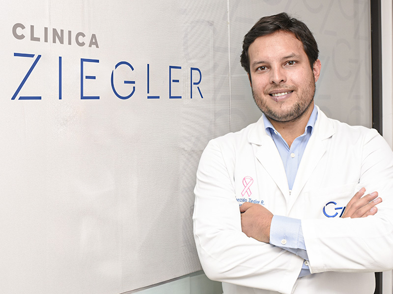
Interview with Dr. Gonzalo Ziegler
(Lima, Peru)
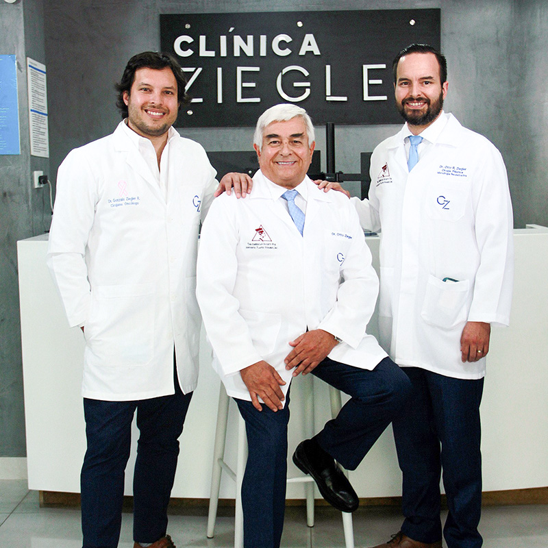
In this interview, Dr. Gonzalo Ziegler, leading expert and director of the renowned clinic “Clinica Ziegler” in Lima, which he runs together with his father and brother, shares his profound knowledge in the field of Dermoscopy and Total Body Mapping. He not only provides insights into his workflow and the role FotoFinder plays in his clinic, but also shares personal anecdotes and valuable feedback from his patients.
FF Why is skin cancer awareness so important?
Dr. Gonzalo Ziegler Skin cancer is one of the most common cancers in the world, mainly due to sun exposure. So, if you are exposed to the sun in childhood, adolescence or at work, even a single sunburn or reddening of the skin can be enough to cause skin damage later in life.
FF How does FotoFinder's technology help with early detection?
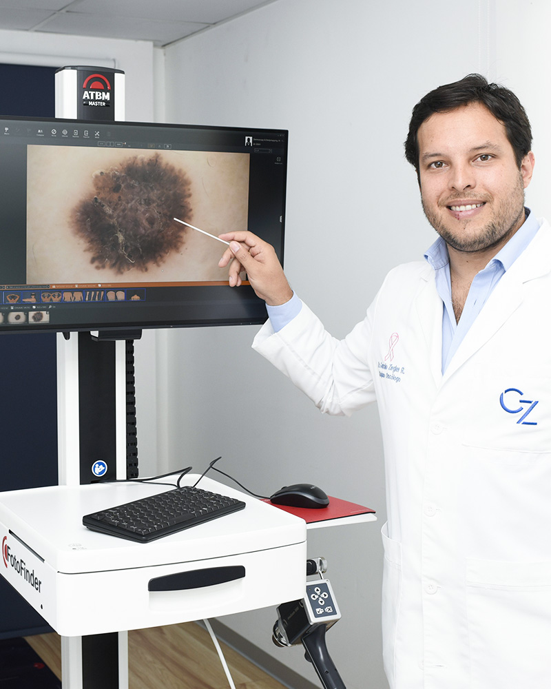
Dr. Gonzalo Ziegler FotoFinder's digital Dermoscopy and Total Body Mapping technology helps us to differentiate between changed and unchanged lesions or if there are signs of a melanoma. This technology has helped us a lot in differentiating between melanocytic and non-melanocytic lesions, including the detection of small skin tumors.
We had the opportunity to diagnose and treat a very well-known person. Her dermatologist had recommended a biopsy for a mole on her breast. Using FotoFinder, we examined her entire body and found that the mole on her breast was harmless. However, we noticed a change on her upper lip. She mentioned that it was a wart that her dermatologist had been treating with laser therapy for the past five years. After dermoscopy, we decided to remove the conspicuous mole. It turned out to be the smallest basal cell carcinoma I have ever seen.
A dedicated team worked to ensure that this person, who depends on her appearance and gives daily television interviews, looks as good as before. After three months of treatment, she now regularly posts Instagram stories and selfies. You can no longer see where the skin cancer was. That's the beauty of our work, especially in combination with the FotoFinder device, which effectively supports us in our daily practice.
FF Can you tell us more about your clinic and how you treat mole patients?
Dr. Gonzalo Ziegler A few years ago, during the pandemic, we experienced a story that led to the founding of our specialty clinic. A patient came to us with a scar on his face after a fall in the shower and asked my father and brother for help. During the examination, he also mentioned a swelling in his armpit. My brother, a surgical oncologist, discovered a conspicuous mass. It turned out to be a metastatic melanoma that had developed from a mole treated with a laser three years ago but not sent to pathology.
Unfortunately, the patient died within a year due to the melanoma. This experience revealed the problems of dealing with skin cancer and melanoma, especially in our country where the healthcare system is often inadequate. As a truly private clinic with no collaboration with insurance companies, we focus on providing patients with the best possible care.
We decided to develop a structured approach to patient care. The first step is a thorough skin examination, which we defined as a specific consultation. Every person requesting a check-up receives a detailed examination by a surgical oncologist, during which the entire skin is carefully examined.
FF How do you usually approach diagnosis and treatment in your clinic?
Dr. Gonzalo Ziegler Any skin change that seems suspicious or worries the patient will be marked. If skin cancer is suspected, the case is given the highest priority and we immediately take dermoscopic images with FotoFinder. Our aim is to resolve such cases within a week. Patients with lower priority are scheduled for a FotoFinder examination within a week and receive their final report within a month.
In around 95% of high-priority patients, we proceed with surgery and complete the treatment within three months. This is crucial because we don't let patients with suspicious lesions leave without action. They see the images of their skin and often want to do something, especially if the skin is damaged or the lesion is unsightly. Then we perform a small biopsy to confirm the diagnosis. Afterwards we make a detailed treatment plan that considers both oncological and cosmetic aspects. This may include surgical removal and, if required, a sentinel lymph node biopsy. Our results are very good. Even melanomas in the advanced stages III or IV, we can treat with immunotherapies and medication.

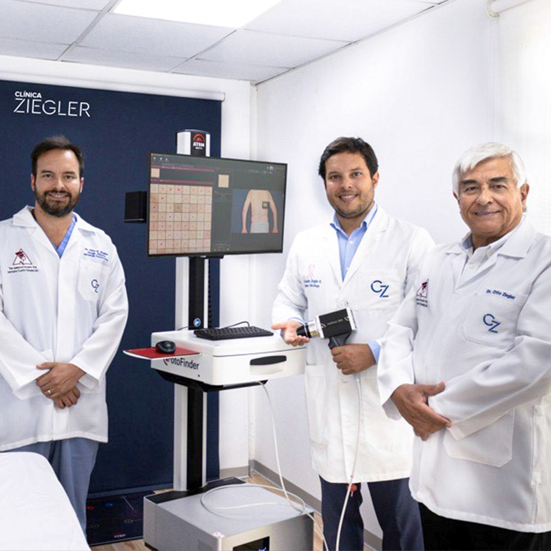
FF Why are Total Body Mapping and dermoscopy with FotoFinder crucial for early detection?
Dr. Gonzalo Ziegler Our goal is to prevent, diagnose and treat diseases at an early stage. In this context, FotoFinder has taken a big step forward in closing this gap. Before the introduction of FotoFinder, the number of patients who consulted us was lower and the time from diagnosis to treatment was longer.
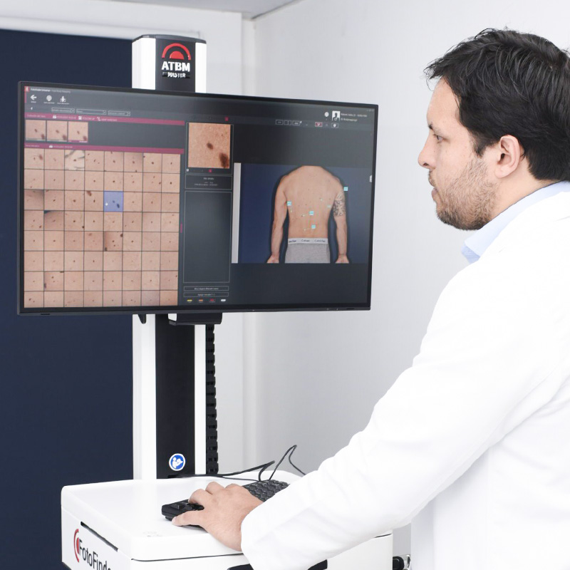
On the large monitor, which has practically the size of a television, we can enlarge skin lesions and measure them using artificial intelligence. This technology allows us to switch between different light modes and carry out a detailed analysis. Patients are impressed by the examination, as such technology did not exist a few years ago. In the early stages of the disease, high-quality dermatoscopic images are often more valuable than blood tests, CT scans or PET scans.
FF What is the success story of your clinic?
I think the success of our skin cancer clinic is the fact that we have embraced this technology. Many colleagues in dermatology reject modern technology, even though it makes many things easier. Technology will not replace us; we are the ones who use it to improve the treatment for our patients.
FF What feedback do you receive from patients about Total Body Mapping?
When we use the technology with patients, they are often very satisfied and grateful. When we suggest Total Body Mapping, they often ask if this technology is reliable. To reassure them, we show research papers, examples from other patients and photos. The examination can take between 20 and 60 minutes, depending on the number of moles. In our workflow, a technician works with the device all day and analyzes the images. I review the dermoscopy twice: once during the examination and a second time afterwards. I describe each image in detail before the work is considered complete. Because some melanomas are difficult to diagnose, we take the time to review the images thoroughly. Therefore, we prioritize patients in triage according to urgency to ensure that each case is treated carefully.