



After the opulent festivities, Sherlock plays his violin and hangs out with his thoughts.

“What are you doing? Why don't you help me - the guests are about to arrive for the New Year's Eve soirée.”

“I am poring over the case of Professor Blum from Constance, which came by dispatch yesterday. Very exciting....”
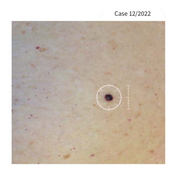
“A 54-year-old female patient with a dark nodular lesion on the left shoulder, 8x15mm in size, new and rapidly growing.”
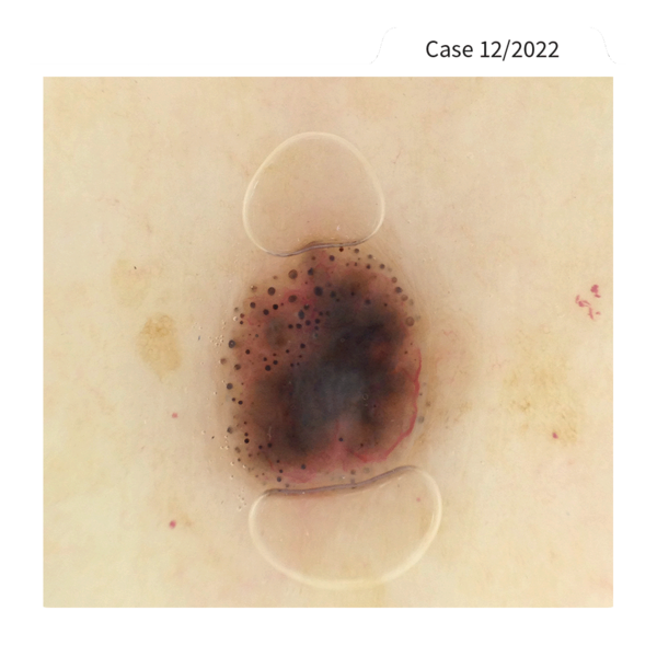
“Professor Blum took a closer look with the handyscope: eccentric homogeneous dark dots and globules, discrete white lines at the edge in the lower center ... Overall brown and blue-gray colors. What could this be? Melanoma? And if not, what else? Hmmm ....”

“Doubts, my dear Sherlock? Well, there is only one solution!”

“Oh yeah? And would you mind telling me which one it is? Or do you want to wait until the New Year to reveal the secret?”

“Well, it's quite simple: if you can't make a definite diagnosis, excision is a MUST. It is hard to imagine what can happen if you are wrong and a supposedly harmless lesion turns out to be melanoma ...”

“Very good, Dr. Watchon. Let's wait and see what conclusion Professor Blum comes to – do you hear knock on the door, too, Watchon? So early?"

“This early? You're kidding, Moles! If you'd helped me set the table.... Anyway, our guests are already waiting.”

“Welcome to 221B Baker Street, Professor Mohariarty!”

“Mr. Waker - I'm glad you accepted our invitation. Come in!”

“You invited Mohariarty and Waker to celebrate New Year's Eve with us??? The very two who last challenged me and tried to play me foul?!”

“Come on, Sherlock. Despite all the competition, we are all part of the FotoFinder Family and dedicated to the well-being of skin cancer patients. But - there's still someone missing ...”

“Ah, there you are, Professor Blum. Come in - I think someone will be very pleased to meet you!”

“Professor Blum? But how ...? Well, this is a surprise! Watchon and I were just talking about your case. Watchon concluded that surgery and histology were required in this case. And you ...?”

“That's exactly what we did, Mr. Moles! If you can't classify a lesion as benign with absolute certainty, the management decision must be: Excision and histology!
It was a pigmented basal cell carcinoma, by the way.”

“This means that pigmented basal cell carcinoma can simulate melanoma! Astounding! So this case is also solved, just in time for the end of what has been a very adventurous year. May I toast to this with all of you? ...”

Sherlock's toast is abruptly interrupted by the ringing of church bells. It is midnight. The turn of the year.

“Let's toast to the NEW YEAR! I wish us all one thing above all: Health! For us and especially for the people we help to stay or become healthy. That's what connects us all.“

“To the great dermoscopy community”


Thank you for following the adventures of Sherlock Moles and Dr. Watchon. Our illustrator was able to accompany them for a year. Will the paths of the three cross again? Let us surprise you ...




Sherlock is out and about in London very late at night ... or rather very early: it's already dawn when he arrives at 221B Baker Street.
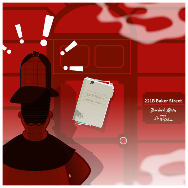
At the front door, he finds an alarming message....
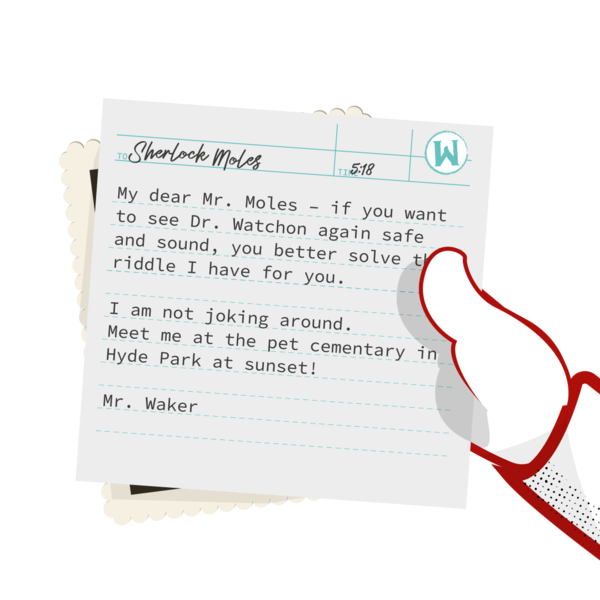
"If you want to see Dr. Watchon again, solve the riddle of Dr. Rosario Peralta - then nothing will happen to your colleague. Sincerely, Mr. Waker!"
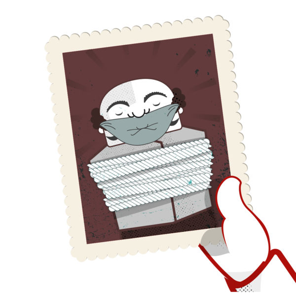
"Oh my goodness, what have they done to him?"

"Don't worry, Watchon, I'll get you out of this! Whoever Mr. Waker is – he will get a nasty surprise!"
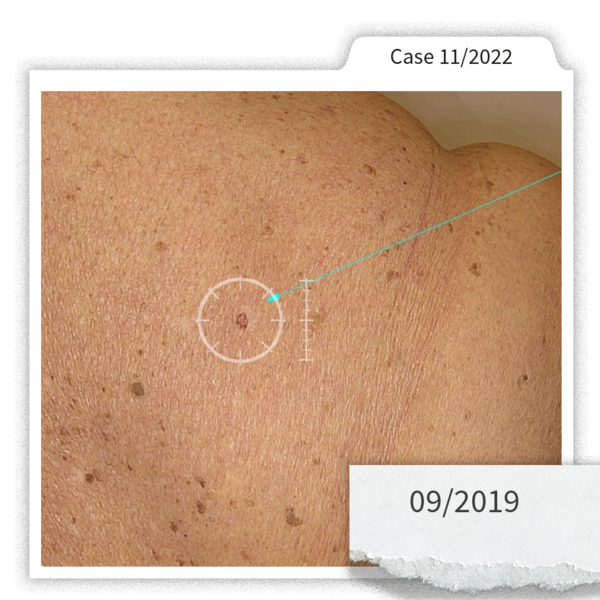
Dr. Peralta's note: "In August 2019, a 76-year-old woman with personal history of in-situ superficial spreading melanoma on her left leg (1999) came for a skin check. Total body exam was performed using the vexia system and medicam 1000. Clinically, she presented a scar lesion with brown irregular pigment on her back."
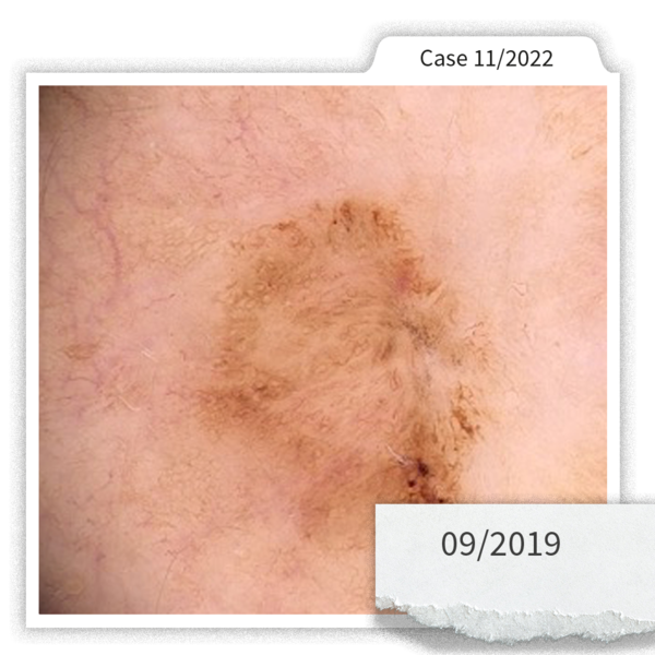
"The patient said that the scar was secondary to a shave biopsy in 2017 which anatomopathologically revealed a dysplastic nevus. Dermoscopic examination showed brown pigmentation with a chaotic growth pattern beyond the scar’s edge. Wiht the suspected of recurrent melanoma, excisional biopsy was indicated."
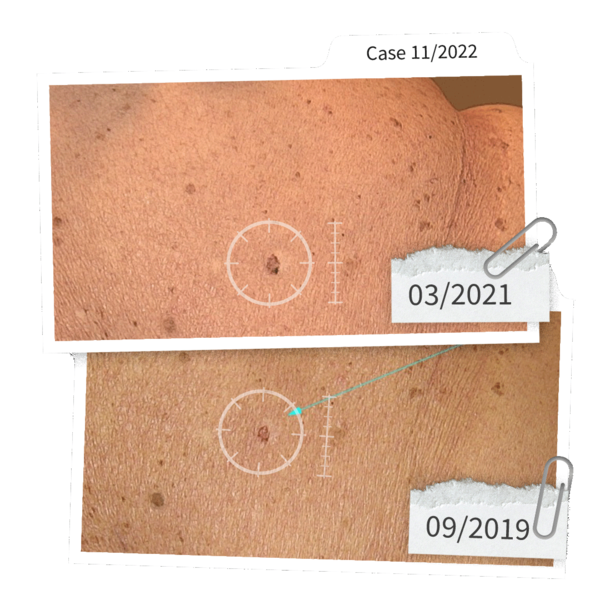
"Second visit in March 2021. After the first visit, the patient had not had the lesion removed; she had cancelled all medical appointments for fear of SARS-COV 2. Clinical comparative exam presented a growing, irregularly pigmented lesion."
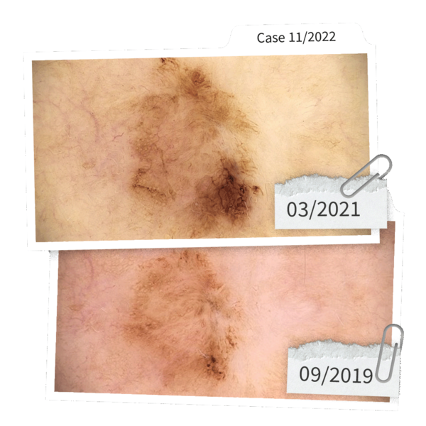
"Examination with medicam 1000 showed brown pigmentation with a chaotic growth pattern beyond the scar’s edge as well as eccentric brown hyperpigmentation with some angulated lines. As a result, the lesion was removed."

"The riddle you have to solve, Mr. Moles: was it a recurrent nevus or recurrent melanoma? Come to the pet cemetery in Hyde Park or your colleague's fate is sealed!"

"Waker – you’ll pay for that! Watchon, I'll get you out of there!"
A little later in Hyde Park.

"How nice that you found your way to us so quickly, dear Sherlock. Lately, you have exposed my colleague Mohariarty (see Episode VII), and now you are going to pay for this! So Mr. Moles - what is the diagnosis? Recurrent nevus or recurrent melanoma?"

“What gain were the clues for recurrent nevus... trauma or surgical-cosmetic procedure .... repigmentation after a few months, confined to the scar.... in dermoscopy, radial lines, symmetry, centrifugal growth pattern..."
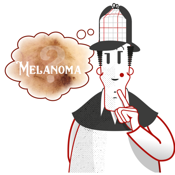
"And the clues for recurrent melanoma .... also trauma or surgical-cosmetic intervention .... repigmentation after a year, beyond the scar’s edge with extension to normal skin – this is the strongest clue for melanoma – in dermoscopy, circles, eccentric hyperpigmentation at the periphery, chaotic, non-continuous growth pattern, pigmentation beyond the scar’s edge....”

"All indications point to recurrent melanoma. The biopsy certainly confirmed this!"

"You are amazing, Mr. Moles. That was really well deduced! But I'm a man of my word. Here you have Dr. Watchon back..."
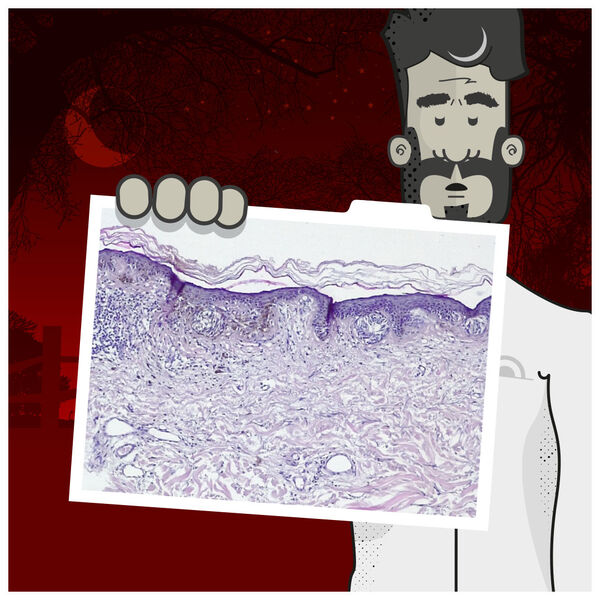
"Dr. Peralta came to the same conclusion. And histology clearly shows you were both right! The anatomopathological result revealed a lentigo maligna melanoma ( in-situ melanoma) near the scar…"

"Thanks Sherlock. That was a last second rescue! Mr. Waker has already bragged that if you fail, you should only be called 'Sherlock Guaca-Moles'..."

"Hahaha, guaca-moles isn't bad, I'll have to remember that one. How lucky that I don't have to be told that...".


You want to share your case in the "Adventures of Sherlock Moles & Dr. Watchon"?
Send us your outstanding case with a description and the clinical as well as the dermoscopy pictures tomarketingfotofinder.de!

![[Translate to English:] FotoFinder Systems, Sherlock Moles, Dr. Watchon, Case of the month, Ana Sortino](/fileadmin/_processed_/d/2/csm_FF_SherlockMoles_2022-01--Web02_f478d9aaa9.png)
![[Translate to English:] FotoFinder Systems, Sherlock Moles, Dr. Watchon, Case of the month, Ana Sortino](/fileadmin/_processed_/b/9/csm_FF_SherlockMoles_2022-01--Web03_8777edcb20.jpg)
Midnight in foggy London – a dark figure approaches 221B Baker Street.
![[Translate to English:] FotoFinder Systems, Sherlock Moles, Dr. Watchon, Case of the month, Ana Sortino](/fileadmin/_processed_/f/1/csm_FF_SherlockMoles_2022-01--Web04_732fbae922.jpg)
„Come on, Watchon, have a drink with me. If there's nothing exciting happening in this town, we can at least have a glass of wine. – Oh, there is a knock at the door! Are you expecting anyone this late?“
„Certainly not! But anyone who rings this late is sure to bring something interesting...“
![[Translate to English:] FotoFinder Systems, Sherlock Moles, Dr. Watchon, Case of the month, Ana Sortino](/fileadmin/_processed_/4/4/csm_FF_SherlockMoles_2022-01--Web05_1916992e1f.jpg)
„Good Golly, Sherlock, it is …”
![[Translate to English:] FotoFinder Systems, Sherlock Moles, Dr. Watchon, Case of the month, Ana Sortino](/fileadmin/_processed_/3/d/csm_FF_SherlockMoles_2022-01--Web06_3298871a0e.jpg)
„Good evening, Sherlock. Your favorite nemesis is here: Professor MOHAriarty!“
![[Translate to English:] FotoFinder Systems, Sherlock Moles, Dr. Watchon, Case of the month, Ana Sortino](/fileadmin/_processed_/b/c/csm_FF_SherlockMoles_2022-01--Web07_d8d90ec98c.jpg)
„You do claim to be the greatest Mole detective of all time. HA! Then explain this case of Dr. Ana Maria Sortino from São Paulo!”
![[Translate to English:] FotoFinder Systems, Sherlock Moles, Dr. Watchon, Case of the month, Ana Sortino](/fileadmin/_processed_/a/7/csm_FF_SherlockMoles_2022-01--Web08_47ec288107.png)
„In 2018, this 61-year-old patient was referred to Dr. Sortino: Fitzpatrick phototype I, severely sun-damaged skin, family history of melanoma as well as a recent personal history of a thin invasive melanoma on his thigh (Breslow 0.6 mm). Initial Total Body Mapping with the ATBM II system showed a linear excision scar surrounded by solar lentigines and small flat lesions. Over the 4-year-follow-up, multiple BCCs and SCCs were diagnosed and treated.“
![[Translate to English:] FotoFinder Systems, Sherlock Moles, Dr. Watchon, Case of the month, Ana Sortino](/fileadmin/_processed_/1/5/csm_FF_SherlockMoles_2022-01--Web09_5f4f2c4a2a.png)
„In 2021, Total Body Mapping visualized a small lesion with central hyperpigmentation above the scar of the previous melanoma: a macular asymmetric mole with two shades of brown.”
![[Translate to English:] FotoFinder Systems, Sherlock Moles, Dr. Watchon, Case of the month, Ana Sortino](/fileadmin/_processed_/4/c/csm_FF_SherlockMoles_2022-01--Web10_ce95762d7a.jpg)
„And why do you think she noticed that tiny little thing, my dear Sherlock? You usually know everything, har har har.”
![[Translate to English:] FotoFinder Systems, Sherlock Moles, Dr. Watchon, Case of the month, Ana Sortino](/fileadmin/_processed_/c/d/csm_FF_SherlockMoles_2022-01--Web11_54c496c328.png)
„That's easy to explain when you look at the total body images taken in 2018 and 2022: The technology has made a huge leap! The ATBM master, which Dr. Sortino has been using since 2021, allows for brilliant polarized images and thus better assessment of clinical images. So you see, Watchon ... this case of Mohariarty was not such a big mystery either...“
![[Translate to English:] FotoFinder Systems, Sherlock Moles, Dr. Watchon, Case of the month, Ana Sortino](/fileadmin/_processed_/c/c/csm_FF_SherlockMoles_2022-01--Web12_9652616576.png)
„But what kind of lesion are we finally talking about?“
![[Translate to English:] FotoFinder Systems, Sherlock Moles, Dr. Watchon, Case of the month, Ana Sortino](/fileadmin/_processed_/5/a/csm_FF_SherlockMoles_2022-01--Web13_048500ff1a.png)
„The first video dermoscopy image from 2018 revealed central irregular hyperpigmentation surrounded by tan areas. Follow-up serial dermoscopy showed an enlarging lesion with new dermoscopic structures (central atypical pigment network and peripheral granularity, gray dots, and irregular brown circles).“
![[Translate to English:] FotoFinder Systems, Sherlock Moles, Dr. Watchon, Case of the month, Ana Sortino](/fileadmin/_processed_/6/2/csm_FF_SherlockMoles_2022-01--Web14_7e6ea33a4a.png)
„...The documentation also included a report from Moleanalyzer pro, which was used as an additional tool. The Artificial Intelligence Score had moved from the yellow to the gray area, which increased the suspicion of malignancy. A full excision with adequate margins was performed. The final histology report gave the diagnosis of a small diameter (<4mm) MELANOMA IN SITU, superficial spreading type, associated with a dysplastic junctional melanocytic nevus.“
![[Translate to English:] FotoFinder Systems, Sherlock Moles, Dr. Watchon, Case of the month, Ana Sortino](/fileadmin/_processed_/2/d/csm_FF_SherlockMoles_2022-01--Web15_bd53b0fd54.png)
„This case highlights the importance of Total Body Mapping for high-risk patients. Using this method, Ana Maria Sortino was able to clearly detect the change.“
![[Translate to English:] FotoFinder Systems, Sherlock Moles, Dr. Watchon, Case of the month, Ana Sortino](/fileadmin/_processed_/b/8/csm_FF_SherlockMoles_2022-01--Web16_0c7dd64a65.png)
„So no need to worry - we are still the greatest Mole Detectives in the world! And this Mohariarty should look elsewhere, theeheehee...“
![[Translate to English:] FotoFinder Systems, Sherlock Moles, Dr. Watchon, Case of the month, Ana Sortino](/fileadmin/_processed_/9/d/csm_FF_SherlockMoles_2022-01--web17_9fbc5051a9.png)
You want to share your case in the "Adventures of Sherlock Moles & Dr. Watchon"?
Send us your outstanding case with a description and the clinical as well as the dermoscopy pictures to marketingfotofinder.de!




After their holiday, the two Mole detectives walk in idleness in London's Baker Street. Sherlock Moles plays the violin while Dr Watchon immerses himself in his reading.

"Sherlock - you should take a look at this editorial on Assoc. Prof. Zoe Apalla from Greece! She had a tough nut to crack…."

"A case that has already been solved? I'm not really interested in that."
"THAT will interest you, Sherlock. The way the case was solved is remarkable!"
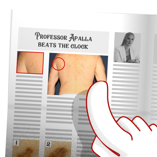
"It's about a 70-year-old man with multiple lesions. Regular check-ups, and a melanoma was already removed 6 years ago."

"During a routine examination, a mole was discovered that showed a slight asymmetrical growth, while all other lesions were unchanged."

"With this amount of moles, ONE nevus was discovered that had only changed slightly? That borders on witchcraft! Let me see..."
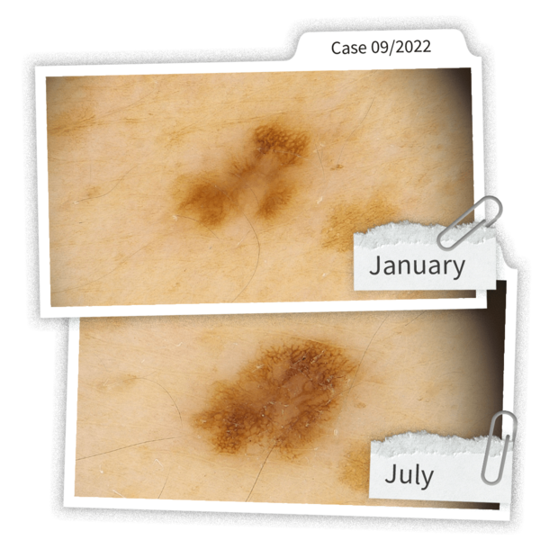
"Above, you see the image from 6 months ago - below the current one. Clearly recognizable, Sherlock! By the way, it is an in situ melanoma."

"That’s true, the change is recognizable thanks to the dermoscopic image, dear Watchon. But how did Prof. Apalla find this one among all the other moles? THAT is the question! Does the article say anything about that?"

"Wait a minute..." *rustle* *rustle*
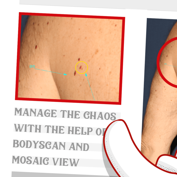
"The article says the search was quite simple - using something called a Bodyscan, which automatically compares full-body images with previous pictures and marks changes. It also says the whole thing only took a few minutes."

“HOLY MOLEY! A computer figured this out so quickly? I tell you, dear Dr. Watchon, if this keeps up, we'll both be out of a job soon!"

"Well, it's not that simple. This is a brilliant visual aid, but Prof. Apalla still had to check it herself and, above all, draw the right conclusions..."


You want to share your case in the "Adventures of Sherlock Moles & Dr. Watchon"
Send us your outstanding case with a description and the clinical as well as the dermoscopy pictures to marketingfotofinder.de!

![[Translate to English:] FotoFinder Systems, Sherlock Moles, Dr. Watchon, Case of the month, Raimonds Karls](/fileadmin/_processed_/9/5/csm_FF_SherlockMoles_2022-01--Web01_d349458474.png)
![[Translate to English:] FotoFinder Systems, Sherlock Moles, Dr. Watchon, Case of the month, Raimonds Karls](/fileadmin/_processed_/f/c/csm_FF_SherlockMoles_2022-01--Web02_c3d888f25e.png)
![[Translate to English:] FotoFinder Systems, Sherlock Moles, Dr. Watchon, Case of the month, Raimonds Karls](/fileadmin/_processed_/e/7/csm_FF_SherlockMoles_2022-01--Web03_a224c16977.png)
"Come, my dear Watchon – we are going to see Dr. Raimonds Karls in Riga! He wants to show us something special."
![[Translate to English:] FotoFinder Systems, Sherlock Moles, Dr. Watchon, Case of the month, Raimonds Karls](/fileadmin/_processed_/6/c/csm_FF_SherlockMoles_2022-01--Web04_33ab0ada1c.png)
"Lots of clouds today, Sherlock. I've never been to Latvia, by the way. They drink something called Black Balsam there, I've heard."
![[Translate to English:] FotoFinder Systems, Sherlock Moles, Dr. Watchon, Case of the month, Raimonds Karls](/fileadmin/_processed_/8/2/csm_FF_SherlockMoles_2022-01--Web05_b69a38ccf3.png)
"When the case is solved, Watchon. While we're on the way, we can get familiar with it..."
![[Translate to English:] FotoFinder Systems, Sherlock Moles, Dr. Watchon, Case of the month, Raimonds Karls](/fileadmin/_processed_/6/b/csm_FF_SherlockMoles_2022-01--Web06_922899e29e.png)
"We are talking about a 24-year-old man, skin type II, with multiple nevi. No personal or family history of melanoma. Light peripheral rim around some lesions. He went to see Dr. Karls because he noticed something odd."
![[Translate to English:] FotoFinder Systems, Sherlock Moles, Dr. Watchon, Case of the month, Raimonds Karls](/fileadmin/_processed_/7/d/csm_FF_SherlockMoles_2022-01--Web07_bd7bc0a29d.png)
"A lesion on the left side of the anterior surface of the chest that was previously inconspicuous, but suddenly showed a dark pigment."
![[Translate to English:] FotoFinder Systems, Sherlock Moles, Dr. Watchon, Case of the month, Raimonds Karls](/fileadmin/_processed_/6/3/csm_FF_SherlockMoles_2022-01--Web08_cf5844dee6.png)
"Hmmm. I'm curious to hear what Dr. Karls has to say about this..."
![[Translate to English:] FotoFinder Systems, Sherlock Moles, Dr. Watchon, Case of the month, Raimonds Karls](/fileadmin/_processed_/f/4/csm_FF_SherlockMoles_2022-01--Web09_098f031eeb.png)
"Ah, beautiful Riga! Let's go to Dr. Karl's institute - he is already waiting for us."
![[Translate to English:] FotoFinder Systems, Sherlock Moles, Dr. Watchon, Case of the month, Raimonds Karls](/fileadmin/_processed_/a/7/csm_FF_SherlockMoles_2022-01--Web10_ea3f58f351.png)
“Mr. Moles, Dr. Watchon – glad you could accept my invitation."
![[Translate to English:] FotoFinder Systems, Sherlock Moles, Dr. Watchon, Case of the month, Raimonds Karls](/fileadmin/_processed_/f/d/csm_FF_SherlockMoles_2022-01--Web11_b5c5887c60.png)
“The patient’s first visit to the clinic was in September 2021. At that time, images of polarized and non-polarized dermatoscopy were taken. The purpose of this two-light examination is to be able to identify white streaks, so-called chrysalis-like structures.”
![[Translate to English:] FotoFinder Systems, Sherlock Moles, Dr. Watchon, Case of the month, Raimonds Karls](/fileadmin/_processed_/2/6/csm_FF_SherlockMoles_2022-01--Web12_3e5c24ebc9.png)
"The FotoFinder AI Score for this lesion was 0.93, quite high!"
![[Translate to English:] FotoFinder Systems, Sherlock Moles, Dr. Watchon, Case of the month, Raimonds Karls](/fileadmin/_processed_/a/2/csm_FF_SherlockMoles_2022-01--Web13_91dfaf28aa.png)
"So you recommended a biopsy, I assume."
![[Translate to English:] FotoFinder Systems, Sherlock Moles, Dr. Watchon, Case of the month, Raimonds Karls](/fileadmin/_processed_/4/9/csm_FF_SherlockMoles_2022-01--Web14_ac90982ee5.png)
"Exactly! The only problem was that the patient didn't come back until February 2022, five months later."
![[Translate to English:] FotoFinder Systems, Sherlock Moles, Dr. Watchon, Case of the month, Raimonds Karls](/fileadmin/_processed_/8/4/csm_FF_SherlockMoles_2022-01--Web15_ad33116446.png)
"On clinical examination, the lesion showed a progressive growth of dark structureless fields without significant dynamics, but dermatoscopically over 156 days progression of black structureless areas. Observing these changes, the patient agreed to a biopsy of the lesion."
![[Translate to English:] FotoFinder Systems, Sherlock Moles, Dr. Watchon, Case of the month, Raimonds Karls](/fileadmin/_processed_/0/0/csm_FF_SherlockMoles_2022-01--Web16_60a40d2e57.png)
"Histology revealed that the lesion was a combined Spitz nevus, a rare type of the Spitz family lesions."
![[Translate to English:] FotoFinder Systems, Sherlock Moles, Dr. Watchon, Case of the month, Raimonds Karls](/fileadmin/_processed_/5/6/csm_FF_SherlockMoles_2022-01--Web17_65aebc3941.png)
"So the Artificial Intelligence Score was wrong?"
![[Translate to English:] FotoFinder Systems, Sherlock Moles, Dr. Watchon, Case of the month, Raimonds Karls](/fileadmin/_processed_/1/3/csm_FF_SherlockMoles_2022-01--Web18_2c1dffc429.png)
"I wouldn't say that. Usually, the AI is not trained on such rare lesions – but it clearly indicated that I should take a closer look, which was absolutely correct. But in the end, the doctor makes the diagnosis!"
![[Translate to English:] FotoFinder Systems, Sherlock Moles, Dr. Watchon, Case of the month, Raimonds Karls](/fileadmin/_processed_/9/1/csm_FF_SherlockMoles_2022-01--Web19_4926b18c8b.png)
"Thank you, dear Dr. Karls – that was really a special case and worth the trip. But now we would like to try this Black Balsam, my friend!"
![[Translate to English:] FotoFinder Systems, Sherlock Moles, Dr. Watchon, Case of the month, Raimonds Karls](/fileadmin/_processed_/2/6/csm_FF_SherlockMoles_2022-01--web20_9b787af529.png)
![[Translate to English:] FotoFinder Systems, Sherlock Moles, Dr. Watchon, Case of the month, Raimonds Karls](/fileadmin/_processed_/e/c/csm_FF_SherlockMoles_2022-01--web21_be92719c44.png)
You want to share your case in the "Adventures of Sherlock Moles & Dr. Watchon"
Send us your outstanding case with a description and the clinical as well as the dermoscopy pictures to marketingfotofinder.de!

![[Translate to English:] FotoFinder Systems, Sherlock Moles, Dr. Watchon, Case of the month, Michaela Starace](/fileadmin/_processed_/9/9/csm_FF_SherlockMoles_2022-01--Web01_051ac0fd06.png)
![[Translate to English:] FotoFinder Systems, Sherlock Moles, Dr. Watchon, Case of the month, Michaela Starace](/fileadmin/_processed_/0/d/csm_FF_SherlockMoles_2022-01--Web02_c5831cdcb2.png)
Late breakfast, Sherlock Moles and Dr. Watchon's most important meal, is abruptly interrupted by a knock on the door....
![[Translate to English:] FotoFinder Systems, Sherlock Moles, Dr. Watchon, Case of the month, Michaela Starace](/fileadmin/_processed_/1/e/csm_FF_SherlockMoles_2022-01--Web03_08015bc13e.png)
"Sherlock – an urgent telegram from Bologna with a mysterious case!"
![[Translate to English:] FotoFinder Systems, Sherlock Moles, Dr. Watchon, Case of the month, Michaela Starace](/fileadmin/_processed_/c/a/csm_FF_SherlockMoles_2022-01--Web04_d8be572a1a.png)
+++ Urgent +stop+ Need your assessment +stop+ Strange phenomenon in a 57 year old patient +stop+ Meeting today 11am Crystal Palace +fullstop+ +++
![[Translate to English:] FotoFinder Systems, Sherlock Moles, Dr. Watchon, Case of the month, Michaela Starace](/fileadmin/_processed_/4/6/csm_FF_SherlockMoles_2022-01--Web05_7e88956527.png)
"What might this be all about, Watchon? – Come on, call us a cab!"
![[Translate to English:] FotoFinder Systems, Sherlock Moles, Dr. Watchon, Case of the month, Michaela Starace](/fileadmin/_processed_/a/1/csm_FF_SherlockMoles_2022-01--Web06_8c167dc0be.png)
"That lady there – that must be the enigmatic patient..."
![[Translate to English:] FotoFinder Systems, Sherlock Moles, Dr. Watchon, Case of the month, Michaela Starace](/fileadmin/_processed_/f/9/csm_FF_SherlockMoles_2022-01--Web07_c49c260c43.png)
"Mr. Moles, thank you for coming. I'm desperate. May I show you my fingertip?"
![[Translate to English:] FotoFinder Systems, Sherlock Moles, Dr. Watchon, Case of the month, Michaela Starace](/fileadmin/_processed_/d/d/csm_FF_SherlockMoles_2022-01--Web08_407017a200.png)
"All right. Let’s have a look with the dermatoscope…"
![[Translate to English:] FotoFinder Systems, Sherlock Moles, Dr. Watchon, Case of the month, Michaela Starace](/fileadmin/_processed_/b/f/csm_FF_SherlockMoles_2022-01--Web09_12f324db24.png)
"Interesting. A conspicuous discoloration under the nail. – Your expertise, Dr. Watchon?"
![[Translate to English:] FotoFinder Systems, Sherlock Moles, Dr. Watchon, Case of the month, Michaela Starace](/fileadmin/_processed_/1/c/csm_FF_SherlockMoles_2022-01--Web10_0f16fd9de1.png)
"Hmm, hard to say. May I kindly ask you to show me your other hand?"
![[Translate to English:] FotoFinder Systems, Sherlock Moles, Dr. Watchon, Case of the month, Michaela Starace](/fileadmin/_processed_/d/a/csm_FF_SherlockMoles_2022-01--Web11_5ca097a562.png)
"I see. These discolourations occur on all fingertips!"
![[Translate to English:] FotoFinder Systems, Sherlock Moles, Dr. Watchon, Case of the month, Michaela Starace](/fileadmin/_processed_/5/2/csm_FF_SherlockMoles_2022-01--Web12_0afae1a7f5.png)
"I may have an explanation for this. Without wanting to be indiscreet, Madam: Did anything change in your life just prior to these discolorations?
"Yes, something did indeed happen – Some time ago I was diagnosed with breast cancer, which is being treated with chemotherapy. Before that, I didn't have these discolorations..."
![[Translate to English:] FotoFinder Systems, Sherlock Moles, Dr. Watchon, Case of the month, Michaela Starace](/fileadmin/_processed_/1/1/csm_FF_SherlockMoles_2022-01--Web14_0487eacba6.png)
"That's it! The discoloration was caused by the therapy. We call this a drug-induced melanocyte activation."
![[Translate to English:] FotoFinder Systems, Sherlock Moles, Dr. Watchon, Case of the month, Michaela Starace](/fileadmin/_processed_/1/6/csm_FF_SherlockMoles_2022-01--Web15_15a17c8d3a.png)
"Oh! Thank you so much. Now I finally know what I'm dealing with. Will it stay that way?"
![[Translate to English:] FotoFinder Systems, Sherlock Moles, Dr. Watchon, Case of the month, Michaela Starace](/fileadmin/_processed_/3/e/csm_FF_SherlockMoles_2022-01--web16_b35f4ddf3e.png)
"It is to be expected that your nails will recover after the chemotherapy. All the best, Madam!"

You want to share your case in the "Adventures of Sherlock Moles & Dr. Watchon"
Send us your outstanding case with a description and the clinical as well as the dermoscopy pictures to marketingfotofinder.de!
![[Translate to English:] FotoFinder Systems, Sherlock Moles, Dr. Watchon, Case of the month, Gabriel Salerni](/fileadmin/_processed_/e/0/csm_FF_SherlockMoles_2022-01--Web00_5c24bdb4cc.png)
![[Translate to English:] FotoFinder Systems, Sherlock Moles, Dr. Watchon, Case of the month, Gabriel Salerni](/fileadmin/_processed_/a/8/csm_FF_SherlockMoles_2022-01--Web01_37d69bbf51.png)
![[Translate to English:] FotoFinder Systems, Sherlock Moles, Dr. Watchon, Case of the month, Gabriel Salerni](/fileadmin/_processed_/3/2/csm_FF_SherlockMoles_2022-01--Web02_12471c0475.png)
Another tricky case for our two Mole Detectives. This time, Sherlock Moles and Dr. Watchon have a tough nut to crack from Dr. Gabriel Salerni (Rosario, Argentina)…
![[Translate to English:] FotoFinder Systems, Sherlock Moles, Dr. Watchon, Case of the month, Gabriel Salerni](/fileadmin/_processed_/d/f/csm_FF_SherlockMoles_2022-01--Web03_302329d254.png)
Just before the 5 o'clock tea, they notice a strange envelope that had been slipped under their door.
![[Translate to English:] FotoFinder Systems, Sherlock Moles, Dr. Watchon, Case of the month, Gabriel Salerni](/fileadmin/_processed_/5/6/csm_FF_SherlockMoles_2022-01--Web04_d323a9a201.png)
"Oh, my dear Sherlock - you seem to have met your match this time. Impossible to solve... look at THIS!"
![[Translate to English:] FotoFinder Systems, Sherlock Moles, Dr. Watchon, Case of the month, Gabriel Salerni](/fileadmin/_processed_/3/2/csm_FF_SherlockMoles_2022-01--Web05_158cd3c238.png)
"Indeed, my dear Watchon. A lot of moles, in all variations. To grasp that..."
"...should be impossible, shouldn't it, Sherlock?
![[Translate to English:] FotoFinder Systems, Sherlock Moles, Dr. Watchon, Case of the month, Gabriel Salerni](/fileadmin/_processed_/a/e/csm_FF_SherlockMoles_2022-01--Web06_06296cab4b.png)
"My dear Watchon, not impossible at all. All it takes is a thousand eyes!"
![[Translate to English:] FotoFinder Systems, Sherlock Moles, Dr. Watchon, Case of the month, Gabriel Salerni](/fileadmin/_processed_/8/4/csm_FF_SherlockMoles_2022-01--Web07_c42317de48.png)
"A thousand eyes? You must be joking, Sherlock. How do you do that?"
![[Translate to English:] FotoFinder Systems, Sherlock Moles, Dr. Watchon, Case of the month, Gabriel Salerni](/fileadmin/_processed_/7/e/csm_FF_SherlockMoles_2022-01--Web08_409612adb7.png)
"Well, wouldn't it be convenient to have something that detects all lesions and remembers size, pattern and position? Then you would only have to look at the moles that have changed."
![[Translate to English:] FotoFinder Systems, Sherlock Moles, Dr. Watchon, Case of the month, Gabriel Salerni](/fileadmin/_processed_/d/f/csm_FF_SherlockMoles_2022-01--Web09_f53b7b89e7.png)
"Here, for example, this 53-year-old male patient consulted Dr. Salerni because of a very atypical mole. But through the Artificial Intelligence of 1000 Eyes, something completely different was discovered with the ATBM master..."
![[Translate to English:] FotoFinder Systems, Sherlock Moles, Dr. Watchon, Case of the month, Gabriel Salerni](/fileadmin/_processed_/d/2/csm_FF_SherlockMoles_2022-01--Web10_e3577f5582.png)
"... specifically, a fast-growing lesion, a melanoma in situ, which could be removed without any problems thanks to early detection!"
![[Translate to English:] FotoFinder Systems, Sherlock Moles, Dr. Watchon, Case of the month, Gabriel Salerni](/fileadmin/_processed_/a/b/csm_FF_SherlockMoles_2022-01--Web11_117d464d6e.png)
"Can you spot the difference, dear Dr. Watchon?"
![[Translate to English:] FotoFinder Systems, Sherlock Moles, Dr. Watchon, Case of the month, Gabriel Salerni](/fileadmin/_processed_/6/3/csm_FF_SherlockMoles_2022-01--Web12_911dc57be2.png)
"So it's always better to take a second look. Even better: 1000 glances... "
![[Translate to English:] FotoFinder Systems, Sherlock Moles, Dr. Watchon, Case of the month, Gabriel Salerni](/fileadmin/_processed_/0/d/csm_FF_SherlockMoles_2022-01--Web13_661d2bb111.png)

You want to share your case in the "Adventures of Sherlock Moles & Dr. Watchon"
Send us your outstanding case with a description and the clinical as well as the dermoscopy pictures to marketingfotofinder.de!



Dr. Ahmed Sadek from Cairo (Egypt) has approached Sherlock Moles directly with an extraordinary case. The results of his examination have surprised him so much that Sherlock consults his colleague Dr. Watchon...

It is a beautiful spring morning. Dr. Watchon is enjoying the peace and quiet, sitting unsuspectingly in his wing chair and browsing through his favorite morning paper, when suddenly...

... Sherlock Moles enters the room. "Dr. Watchon - I have a very interesting case here from Dr. Sadek.
This 27-year-old patient has had this lesion for only three weeks, and it was painful at the beginning. What do you think of it?"
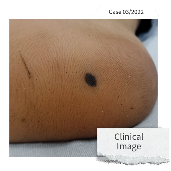
"Hmm, at first glance a harmless birthmark. Let me see the magnification!"
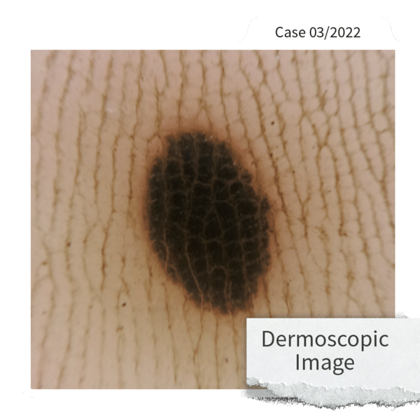
"Hmmm... again - not particularly suspicious. I see a black oval lesion with the medicam 1000s. It appears as if there is a slight red hue peri-lesionally, but..."

"Sherlock, are you kidding me? That looks perfectly normal. This is not a case for you, Mr. Moles..."

"Oh really? Well, take a closer look. Sometimes you have to scratch the surface - literally."
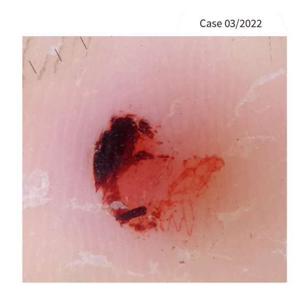
"You see, Watchon? By ablating the surface of the skin, the lesion has almost disappeared, leaving a more distinct reddish coloration that clearly suggests a sub-corneal hematoma."

"A deep-seated hematoma? I would never have guessed THAT after the first two pictures! How did you...?"

"Well, my dear Watchon – sometimes you have to remove the obvious first to get to the bottom of it and then take a deep dermoscopic look beneath the skin!"

Join Sherlock Moles & Dr. Watchon here for a look under the skin of more tricky cases from Dr. Ahmed Sadek!

You want to share your case in the "Adventures of Sherlock Moles & Dr. Watchon"?
Send us your outstanding case with a description and the clinical as well as the dermoscopy pictures to marketingfotofinder.de!


This month, Sherlock Moles and Dr. Watchon will have a look at a Total Body Dermoscopy case from the great Dr. Paweł Pietkiewicz (Poland).
He shared the case of a solitary lesion on the foot which showed changes in the follow-up.
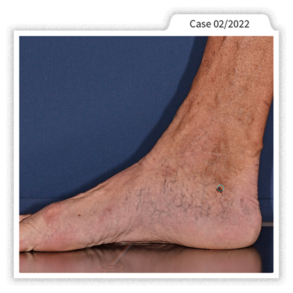
This picture shows the clinical image of the lesion.
The Body Mapping was taken with the ATBM master from FotoFinder.

This is the lesion of a 62-year-old high-risk patient with a history of melanoma and an atypical nevus syndrome with multiple lesions presenting chaotic architecture.
Look at this mole! Very interesting. What do you think?
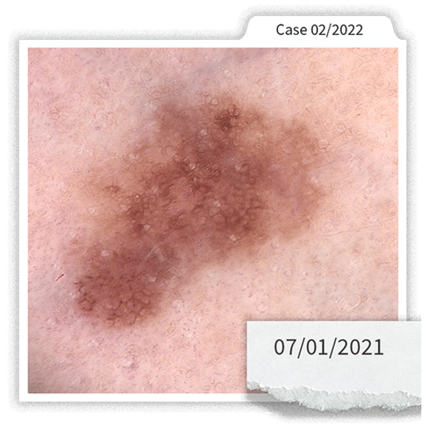
Well, let's take a dermoscopy picture!
Dr. Paweł Pietkiewicz uses a medicam 1000s with D-Scope IV with 20× magnification and decides to follow-up the lesion.
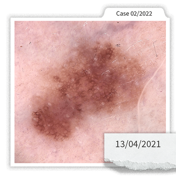
Three months later: A follow-up dermoscopy image was taken with the medicam 1000s and the D-Scope IV and the same magnification.

I can see a slight change at three months follow-up.
But is it because the skin was stretched a little bit?
We should follow this trace for some more months!
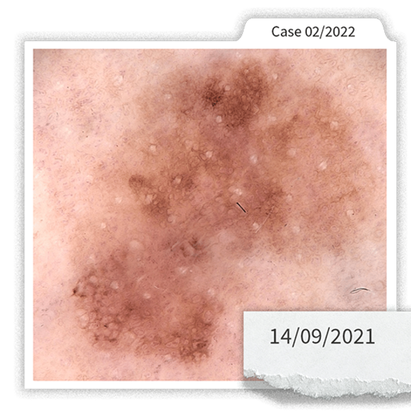
Due to the pandemic the patient sees Dr. Paweł Pietkiewicz after five months, a little farther than he was asked to come for a mole check.
Let's see, what the medicam 1000s will visualize this time!

Dr. Watchon, you were right! Now, the changes are striking!
This is a highly dynamic lesion with clues to malignancy.
It should be excised and sent to histology.

Sherlock Moles and Dr. Watchon received the pathology report.
It was indeed a melanoma, NOS with Breslow thickness: 0.4 mm.


You want to share your case in the "Adventures of Sherlock Moles & Dr. Watchon"?
Send us your outstanding case with a description and the clinical as well as the dermoscopy pictures to marketingfotofinder.de!
-
Episode IXDecember 2022
-
Episode VIIINovember 2022
-
Episode VIIOctober 2022
-
Episode VISeptember 2022
-
Episode VJune 2022
-
Episode IVMay 2022
-
Episode IIIApril 2022
-
Episode IIMarch 2022
-
Episode IFebruary 2022